Friedrich August Kekulé von Stradonitz

Friedrich August Kekulé (Darmstadt, 7 de setembro de 1829 — Bonn, 13 de julho de 1896) foi um químico alemão. Inovou o emprego de fórmulas desenvolvidas em química orgânica, criou em 1857, a Teoria da Tetracovalência do carbono, criou hipótese das ligações múltiplas e propôs, em 1865, após um sonho que teve, a fórmula hexagonal do benzeno.
Friedrich August Kekulé Von Stradonitz nasceu em 7 de setembro de 1829 em Darmstadt, Alemanha. Família descendente de uma linha Tcheca, nobre família da Boêmia. Quando jovem seus hobbies eram caminhadas botânicas, recolhendo e desenhando borboletas. Iniciou seus estudos no ginásio de Darmstadt, sempre um bom aluno com aptidão para línguas, isso resultou na capacidade de falar francês, italiano e inglês, bem como se alemão nativo. Tinha interesse por ginásticas, danças e malabarismo, além do talento em mímicas! Tinha talento para desenho, assim tinha a intenção se tornar um arquiteto. No entanto mal ele sabia o que o destino tinha lhe preparado...
1847
Concluiu seu ginásio e logo ingressou na Universidade de Giessen, no curso de arquitetura; Inscreveu-se em uma aula de química ministrada pelo famoso químico Justus Von Liebig; A partir dessa aula floresceu o interesse pela química; Tomou a decisão de mudar o seu curso de arquitetura e passou a se dedicar ao estudo da química;
1851 - 1852
Teve sua graduação concluída com êxito, mesmo com a não concordância de sua família; Viajou a Paris para continuar seus trabalhos e iniciar seu doutorado; Obteve conhecimentos sobre a teoria unitária da química e teoria dos radicais, após se tornar amigo de Charles Gerhardt e Jean-Baptiste Dumas. Interessou-se pelos problemas da filosofia da química, o que o acompanhou por toda sua vida;
Retornou a Alemanha onde prosseguiu seu doutorado na mesma universidade onde foi graduado, Universidade de Giessen; Começou um pequeno laboratório químico em Heidelberg, utilizando um equipamento muito fino para realizar diversas pesquisas importantes; Em seu doutorado trabalhou como assistente Adolf Von Planta de Reichenau, o que não o deixava feliz, pois para ele estava faltando estimulação intelectual, ficando assim por pouco tempo; Seu ex-professor Liebig o recomendou para uma posição no hospital St. Bartholomew, em Londres, trabalhando com John Stenhouse, onde obteve uma experiência muito importante;
1853 – 1856
Conheceu Alexander Williamson, o qual estendeu as teorias de Charles Gerhardt de explicar como os éteres podem ser derivados da água e o incentivando a introduzir um novo composto, metano ou gás do pântano; Discutiu assuntos interessantes sobre a química, trabalhando na tentativa de classificar os compostos orgânicos por meio de estrutura, esclarecendo a idéia posterior da tetravalência do carbono e da capacidade dos átomos de carbono formarem cadeias; Percebeu que a teoria dos compostos orgânicos não levou em conta a potência específica de combinação ou valências dos átomos específicos; Foi nomeado professor da Universidade de Heidelberg, na Alemanha, onde finalmente estabeleceu a tetravalência do carbono;
1857 – 1859
Recebeu o cargo de professor de química na Universidade de Gent, na Bélgica; Começou a utilizar representações gráficas de moléculas orgânicas, enfatizando natureza tetravalente dos átomos de carbono e sua capacidade de formar cadeias, assim voltando sua atenção para estrutura do benzeno (composto com propriedades em comum que não poderia ser explicado por qualquer teoria), apresentando sua representação hexagonal.
1860 – 1896
Registrou dezenove fórmulas de ácido acético; Foi chamado para Universidade de Bonn; Propôs que o benzeno tinha uma estrutura na qual seis átomos de carbono formavam um anel, com alternância de ligações simples e duplas. No entanto a química do benzeno nem sempre era consistente com esta fórmula estrutural; Para superar este problema sugeriu duas formas de benzeno, em equilíbrio dinâmico, novamente sua teoria mostrou-se parcialmente correta; Fez um discurso em Berlim por ocasião do vigésimo quinto aniversário de seu anuncio da teoria do benzeno, revelando que suas teorias estruturais foram-lhe reveladas através de um sonho...
O sonho de Kekulé

| Eu estava sentado à mesa a escrever o meu compêndio, mas o trabalho não rendia; os meus pensamentos estavam noutro sítio. Virei a cadeira para a lareira e comecei a dormitar. Outra vez começaram os átomos às cambalhotas em frente dos meus olhos. Desta vez os grupos mais pequenos mantinham-se modestamente à distância. A minha visão mental, aguçada por repetidas visões desta espécie, podia distinguir agora estruturas maiores com variadas conformações; longas filas, por vezes alinhadas e muito juntas; todas torcendo-se e voltando-se em movimentos serpenteantes. Mas olha! O que é aquilo? Uma das serpentes tinha filado a própria cauda e a forma que fazia rodopiava trocistamente diante dos meus olhos. Como se se tivesse produzido um relâmpago, acordei;... passei o resto da noite a verificar as consequências da hipótese. Aprendamos a sonhar, senhores, pois então talvez nos apercebamos da verdade." - Augusto Kekulé, 1865.[1] |
Referências
- ↑ BOYD, E.; MORRISON, R. Mol. In:______. Química orgânica. 12. ed. Lisboa: Fundacao Calouste Gulbenkian, 1995. cap. 14.3, p. 701.
Thomas Hunt Morgan


Thomas Hunt Morgan (Lexington (Kentucky), 25 de Setembro de 1866 — Pasadena, 4 de Dezembro de 1945) foi um zoólogo e geneticista estadunidense.
Devido ao seu trabalho, a Drosophila tornou-se um dos principais modelos animais na área da genética. Suas contribuições mais importantes foram para a genética, pelas quais recebeu o Nobel de Fisiologia/Medicina de 1933, por provar que os cromossomas são portadores de genes.
Morgan recebeu o seu bacharelato da Universidade de Kentucky em 1886 e o seu mestrado em 1888. Recebeu o seu doutoramento da Universidade Johns Hopkins em 1890. Ao trabalhar no desenvolvimento embriônico da Drosophila (a mosca da fruta) na Universidade da Columbia, ele ficou interessado em hereditariedade. As teorias de Gregor Mendel tinham sido recentemente redescobertas por volta de 1900 e Morgan estava interessado em testar essas teorias em animais.
Ele começou por tentar criar híbridos da Drosophila, mas não teve sucesso durante dois anos. Finalmente em 1910, ele reparou num mutante macho com os olhos brancos no meio dos machos que normalmente têm os olhos vermelhos. Ele cruzou este macho de olhos brancos com uma fêmea de olhos vermelhos. A sua progenia tinha olhos vermelhos, sugerindo que a característica era recessiva. Morgan deu o nome white ao gene, começando assim a tradição de nomear os genes pelos seus alelos mutantes. À medida que Morgan continuava a cruzar os seus mutantes reparou que apenas os machos exibiam a característica dos olhos brancos. Disto concluiu que 1) alguns traços estão ligados ao sexo; 2) a característica era provavelmente transportada pelos cromossomas sexuais (cromossomas X e Y); e 3) outros genes eram também provavelmente transportados em cromossomas específicos. Ele e os seus estudantes contaram as características de milhares de moscas da fruta e estudaram as suas heranças. Usando a recombinação de cromossomas, formaram um mapa das localizações dos genes no cromossoma. Morgan e os seus estudantes também escreveram o livro The Mechanism of Mendelian Heredity. Morgan transferiu-se para o Instituto de Tecnologia da Califórnia em 1928. Morgan morreu em Pasadena (Califórnia).
Morgan deixou um importante legado na área da genética. Alguns dos estudantes de Morgan da Universidade de Columbia e do Instituto de Tecnologia da Califórnia (Caltech) seguiram a pesquisa e ganharam os seus próprios prémios Nobel, incluindo George Wells Beadle, Edward B. Lewis e Hermann Joseph Muller. Em homenagem a Morgan, a Genetics Society of America condecora anualmente um dos seus membros com a medalha de Thomas Hunt Morgan por contribuições significantes para a ciência genética. O prémio Nobel Eric Richard Kandel escreveu o seguinte sobre Morgan:
"Assim como as descobertas de Charles Darwin sobre a evolução de espécies animais definiram a biologia do século XIX como uma ciência descritiva, as descobertas de Morgan sobre os genes e sua localização nos cromossomos ajudaram a transformar a biologia em uma ciência experimental.".
Theodosius Dobzhansky

Theodosius Hryhorovych Dobzhansky (Ucraniano— Теодосій Григорович Добжанський; algumas vezes alterado para Theodosius Grigorevich Dobzhansky ou Theodore Dobzhansky; Nemyriv, 24 de janeiro de 1900 — San Jacinto, 18 de dezembro de 1975) foi um geneticista e biólogo evolutivo. Dobzhansky nasceu na Ucrânia (então parte do Império Russo), iniciou os seu estudos na Universidade de Kiev e emigrou para os Estados Unidos em 1927[1].
Biografia
Início da vida
Dobzhansky nasceu em 25 de janeiro de 1900 em Nemirov, Ucrânia, então parte do Império Russo. Era filho único, seu pai Grigory Dobzhansky foi um professor de matemática, e sua mãe era Sophia Voinarsky. Em 1910 a família se mudou para Kiev. Durante seu curso de segundo grau noturno, Dobzhansky colecionou borboletas e decidiu se tornar um biólogo. Em 1915 ele encontrou Victor Luchnik que o convenceu a se especializar em besouros. Dobzhansky trabalhou na Universidade de Kiev entre 1917 e 1921, onde estudou até 1924. Ele então se mudou para Leningrado para estudar com Yuri Filipchenko, onde um laboratório de estudos com Drosophila melanogaster já havia sido montado.
Em 8 de agosto de 1924, Dobzhansky casou-se com a geneticista Natalia Sivertzev que havia trabalhado com I. I. Schmalhausen em Kiev. Os Dobzhanskys tiveram uma filha, Sophie, que se casou com o antropologista americano Michael D. Coe.
Este foi um período de grande revolta social na Rússia, com a Primeira Grande Guerra seguida pela Revolução Russa de 1917 e pela Guerra Civil Russa que estabeleceu a União Soviética.
Pesquisa na América
Dobzhansky emigrou para os Estados Unidos em 1927 como um membro do Conselho Internacional de Educação da Fundação Rockefeller chegando a Nova Iorque em 27 de Dezembro. Ele trabalhou com Thomas Hunt Morgan na Universidade de Columbia, que foi um pioneiro no uso de moscas da fruta (Drosophila melanogaster) em experimentos genéticos. Ele seguiu Morgan quando este foi para o Instituto de Tecnologia da Califórnia de 1930 a 1940. Dobzhansky trabalhou com moscas da fruta também fora do laboratório, no campo, tendo descoberto que diferentes variedades regionais de moscas eram mais similares entre si geneticamente do que moscas de outras regiões.
Em 1937 ele publicou um dos maiores trabalhos da síntese evolutiva moderna, a síntese da biologia evolutiva com genética, intitulada Genetics and the Origin of Species, que entre outras coisas definiu a evolução como “uma mudança na frequencia de alelos dentro de um pool gênico”. Também em 1937, ele se tornou um cidadão naturalizado americano. Durante este período ele teve um desentendimento público com um de seus colaboradores, Alfred Sturtevant, baseado em competência profissional.
Dobzhansky retornou a Universidade de Columbia de 1940 a 1962. Ele então se mudou para o Instituto Rockefeller (pouco antes de se tornar Universidade Rockefeller) até sua aposentadoria em 1971.
Doença final e a Luz da Evolução
Em 1 de junho, 1968 foi descoberto que Dobzhansky estava sofrendo de leucemia linfática e lhe foi informado que teria poucos meses de vida. Natalia morreu de trombose coronariana em 22 de fevereiro de 1969. Em 1971 ele se aposentou mas continuou trabalhando como professor emérito, mudando-se para a Universidade da California.
Ele continuou trabalhando e publicou um famoso ensaio anti-criacionista Nothing in Biology Makes Sense Except in the Light of Evolution. Um leal defensor da Evolução Darwiniana, Dobzhansky, de acordo com Francisco Ayala "foi um homem religioso" crente em um Deus criador através da evolução. Sua leucemia tornou-se mais seria no verão de 1975, em 11 de novembro ele fez uma viagem a San Jacinto, California onde ele morreu de insuficiência cardíaca em 18 de dezembro.
Bibliografia
Livros
- Dobzhansky, Th. 1937. Genetics and the Origin of Species. Columbia University Press, New York. (2nd ed., 1941; 3rd ed., 1951)
- The Biological Basis of Human Freedom (1954).
- Dunn, L. C., & Dobzhansky, Th. 1946. Heredity, Race, and Society. The New American Library of World Literature, Inc., New York.
- Dobzhansky, Th. 1955. Evolution, Genetics, & Man. Wiley & Sons, New York.
- Dobzhansky, Th. 1962. Mankind Evolving. Yale University Press, New Haven, Connecticut.
- Dobzhansky, Th. 1967. The Biology of Ultimate Concern. New American Library, New York.
- Dobzhansky, Th. 1970. Genetics of the Evolutionary Process. Columbia University Press, New York.
- Genetic Diversity and Human Equality (1973).
- Dobzhansky, Th., F.J. Ayala, G.L. Stebbins & J.W. Valentine. 1977. Evolution. W.H. Freeman, San Francisco.
- Dobzhansky, Th., 1981. Dobzhansky's Genetics of Natural Populations I-XLIII. R.C. Lewontin, J.A. Moore, W.B. Provine & B. Wallace, eds. Columbia University Press, New York. (reprints the 43 papers in this series, all but two of which were authored or co-authored by Dobzhansky)
Artigos
- Dobzhansky, Th. 1973. "Nothing in biology makes sense except in the light of evolution" The American Biology Teacher 35: (March): 125-129.
- Dobzhansky, T., and O. Pavlovsky. 1957. "An experimental study of interaction between genetic drift and natural selection" Evolution 11: 311-319
Referências
- ↑ Theodosius Dobzhansky (em inglês). Página visitada em 15 de outubro de 2010.
Calvin Bridges


Calvin Blackman Bridges (11 de Janeiro de 1889 - 27 de Dezembro de 1938) foi um cientista dos Estados Unidos da América, conhecidos pelas suas contribuições no campo da Genética, Juntamente com Alfred Sturtevant e Hermann Joseph Muller, Bridges trabalhou na famosa Sala das Moscas de Thomas Hunt Morgan, na Universidade de Columbia.
Bridges escreveu a sua tese de doutoramento sobre a "não-disjunção como prova da teoria da hereditariedade cromossómica". Apareceu como o primeiro artigo da primeira edição da publicação científica Genetics, em 1916.
O seu trabalho com traços ligados ao sexo, na mosca-da-fruta Drosophila melanogaster, sugeriam que os cromossomas continham genes. Mais tarde, Nettie Maria Stephens conseguiu provar essa hipótese, através da análise de cromossomas da mosca-da-fruta. Bridges escreveu dois artigos apresentando a prova.
A contribuição mais conhecida de Bridges, entre os pesquisadores de Drosophila, foi a observação e documentação dos cromossomas politénicos das glandulas salivares de larvas da mosca. Os padrões de bandeamento desses cromossomas são ainda hoje utilizados como marcadores genéticos.
Leitura adicional
- A. H. Sturtevant, A History of Genetics, (Cold Spring Harbor Laboratory Press,2001). ISBN 0-87969-607-9
- E.A. Carlson, Mendel's Legacy: The Origin of Classical Genetics, (Cold Spring Harbor Laboratory Press, 2004). ISBN 0-87969-675-3
- E.A. Carlson, The Gene: A Critical History, (Iowa State Press, 1989). ISBN 0-8138-1406-5
Alfred Henry Sturtevant

Geneticista americano nascido em Jacksonville, Illinois, conhecido pelo trabalho de Ph.D (1913) em que usou os dados de ligação entre genes para lançar as bases da construção dos mapas genéticos, desenvolvendo uma técnica para mapeamento da locação de genes de cromossomos na mosca Drosófila, sob orientação do professor Thomas Hunt Morgan. Foi o mais jovem dos seis filhos do professor de matemática e posteriormente fazendeiro Alfred Henry Sturtevant e de Harriet Evelyn Morse e neto de Julian M. Sturtevant, que fora um graduado da Yale Divinity School e fundador e presidente do Illinois College, onde seu pai foi professor. Educado na escola pública, em Mobile, entrou (1908) para a Columbia University, levado por seu irmão mais velho e professor de línguas antigas na universidade, Edgar, onde se graduou (1912) defendeu seu PhD e publicou o clássico paper. Embora contratado pela Carnegie Institution (1915),continuou pesquisando na Columbia até quando se mudou (1928) para o California Institute of Technology, onde foi professor de genética e de biologia até sua morte, em Pasadena, California. Ele escreveu muitos documentos importantes e livros, inclusive sendo um dos autores de The Mechanism of Mendelian Heredity (1915).
Figura copiada do BIOGRAPHICAL MEMOIRS VOL. 73: http://stills.nap.edu/readingroom/books/bio73h/
William Astbury

William Thomas Astbury (25 de Fevereiro de 1898 — 4 de Junho de 1961) foi um físico e biólogo molecular inglês que fez estudos pioneiros em moléculas biológicas, utilizando a técnica de difração de raios-X. O seu trabalho sobre a queratina providenciou as bases para a descoberta da alfa-hélice por Linus Pauling.
Também estudou a estrutura do ADN em 1937, tendo dado os primeiros passos na elucidação da sua estrutura.
Ligações externas
Maurice Wilkins

Maurice Hugh Frederick Wilkins (Pongaroa, 15 de Dezembro de 1916 — Londres, 5 de Outubro de 2004) foi um fisiologista da Nova Zelândia.
Foi agraciado com o Nobel de Fisiologia/Medicina de 1962, juntamente com Francis Crick e James Watson, por suas contribuições na descoberta da estrutura em dupla hélice do DNA.
Maurice Wilkins, o físico britânico cujos experimentos propiciaram a celebrada descoberta da estrutura do DNA, morreu na terça-feira em um hospital londrino aos 88 anos, informou a King's College, universidade onde ele trabalhava.
Wilkins, que obteve o Nobel de física e medicina em 1962, no mesmo ano que os descobridores da dupla hélice do DNA, Francis Crick e James Watson, morreu amparado por sua família depois de muitos anos dedicados à ciência.
A comunidade científica expressou seu pesar ante o falecimento do físico, que passou sete anos verificando a hipotética estrutura de dupla hélice do DNA construída por seus dois colegas.
Além disso, foram seus experimentos em cristalografía de Raios-X que possibilitaram o descobrimento de Crick e Watson. Wilkins, e depois Rosalind Franklin, demonstraram que os Raios-X podiam ser utilizados para investigar o DNA.
"Maurice Wilkins desempenhou um papel fundamental em um dos descobrimentos mais importantes da história", afirmou Adrian Hayday, professor de Imunobiologia no londrino King's College.
"O êxito de Watson e Crick na elucidação da estrutura do DNA é universalmente conhecido e reconhecido -acrescentou-. Mas sua dependência dos resultados experimentais conseguidos primeiro por Wilkins e depois por Franklin, é freqüentemente esquecida".
Nesta linha, se pronunciaram outros cientistas, que destacaram a modéstia de Wilkins e sua dedicação à ciência.
"Foi um cientista muito importante, que provavelmente não obteve o reconhecimento que merecia por descobrimentos que revolucionaram a ciência", disse Stephen Minger, professor de Ciências Biomédicas na universidade londrina.
Foi Wilkins que obteve pela primeira vez uma imagem de Raio-X do DNA, que ensinou Crick sobre o ácido desoxirribonucleico e quem inspirou Watson.
"Depois, também foi ele quem verificou que a estrutura de dupla hélice era correta", assinalou o cientista Matt Ridley.
Nascido em 1916, Wilkins estudou Física na Universidade de Cambridge, antes de mudar para a de Birmingham.
Durante a segunda Guerra Mundial, trabalhou na Universidade da Califórnia, onde participou do projeto "Manhattan" da bomba atômica.
Depois da guerra, foi professor de física na Universidade escocesa de Saint Andrews -onde estudava o príncipe William da Inglaterra- e em 1963 passou a ensinar na King's College de Londres, onde fez seus importantes experimentos e produziu imagens da forma da molécula do DNA.
O professor Francis Crick, que junto com Watson é celebrado como descobridor do chamado "segredo da vida", morreu de câncer em julho em San Diego, na Califórnia, também aos 88 anos.
Rosalind Franklin
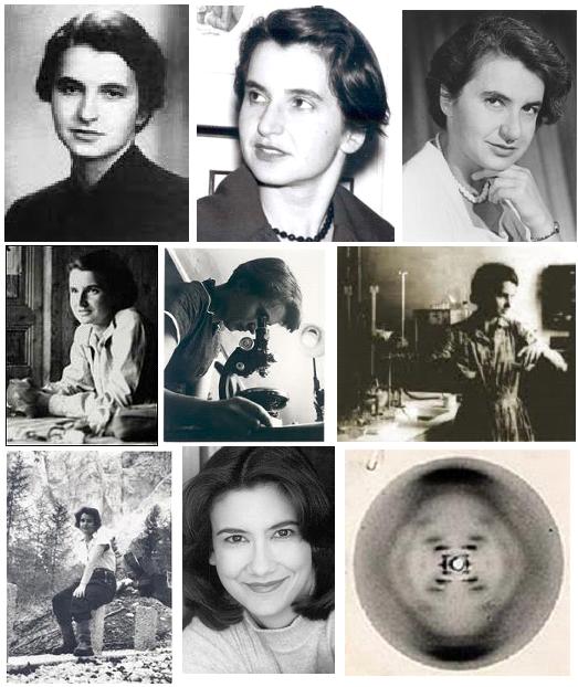

Rosalind Franklin (Londres, 25 de julho de 1920 — Londres, 16 de abril de 1958) foi uma biofísica britânica, pioneira da biologia molecular, que, empregando a técnica da difração dos raios-X, concluiu que o DNA tinha forma helicoidal (1949).[1]
Biografia
Contrariando o desejo dos pais, aos 15 anos ela decidiu que queria ser uma cientista. Entrou (1938) no Newnham College, em Cambridge, graduando-se em físico-química (1941). Iniciou-se como pesquisadora (1942) analisando a estrutura física de materiais carbonizados utilizando raios-X. Trabalhando no British Coal Utilization Research Association, onde desenvolveu estudos fundamentais sobre as microestruturas do carbono e do grafite, base de seu doutorado em físico-química pela Universidade de Cambridge (1945).
Trabalhando em Paris (1947-1950), no Laboratoire Central des Services Chimiques de L'Etat, usou a técnica da difração dos raios-X para análise de materiais cristalinos. Voltando para a Inglaterra, juntou-se a equipe de biofísicos do King's College Medical Research Council (1951) e com Raymond Gosling no laboratório de biofisica do britânico Maurice Wilkins, e iniciou a aplicação de estudos com difração do raio-X para determinação da estrutura da molécula do DNA. Este trabalho permitiu ao bioquímico norte-americano James Dewey Watson e aos britânicos Maurice Wilkins e Francis Crick confirmar a dupla estrutura helicoidal da molécula do DNA, dando-lhes o Nobel de Fisiologia/Medicina (1962), tendo nela a grande injustiçada já que o Nobel não pode ser atribuído postumamente.
Apesar das inúmeras dificuldades provocadas pelo preconceito, ela provou então ser uma cientista de primeiro nível, e mudou-se (1953) para o laboratório de cristalografia J. D. Bernal, do Birkbeck College, em Londres, onde prosseguiu com seu trabalho sobre a estrutura mosaical do vírus do tabaco. Quando iniciou sua pesquisa sobre o vírus da pólio (1956), ela descobriu que estava com câncer. Foi no Birkbeck Collegeaos que publicou seu último trabalho, sobre as estrutura do carvão (1958). Morreu em Londres ainda muito jovem, 37 anos, de câncer no ovário.
Max Delbrück

Max Delbrück (Berlim, 4 de Setembro de 1906 — Pasadena, Califórnia, 9 de Março de 1981) foi um biologista alemão, naturalizado estadunidense.
Max Delbrück nasceu em Berlim, Alemanha. Seu pai era Hans Delbrück, um professor de história na Universidade de Berlim, e sua mãe era a neta de Justus von Liebig. Delbrück estudou astrofísica, e depois física teórica, na Universidade de Göttingen. Depois de receber o seu doutorado em 1930, ele viajou através da Inglaterra, Dinamarca e Suíça. Ele se encontrou com Wolfgang Pauli e Niels Bohr, que o fez se interessar pela biologia. Em 1937, ele mudou-se para os Estados Unidos para prosseguir em seu interesse pela biologia, pesquisando sobre a genética da mosca da fruta Drosophila melanogaster na Divisão de Biologia da Caltech.
Permaneceu nos E.U.A. durante a Segunda Guerra Mundial, ensinando Física na Universidade de Vanderbilt, em Nashville, prosseguindo simultaneamente a sua pesquisa genética. Entre os anos de 1946 e 1953, Max Delbrück integrou temporariamente o grupo reunido sob o nome de Macy Conferences, contribuindo para a consolidação da teoria cibernética.
Em 1969, junto com Alfred Hershey e Salvador Luria, foi agraciado com o Nobel de Fisiologia/Medicina, por investigar e explicar o mecanismo das infecções virais em células vivas.
Faleceu a 9 de março de 1981.
Harvey Itano

Harvey Akio Itano (November 3, 1920 – May 8, 2010) was an American biochemist best known for his work on the molecular basis of sickle cell anemia and other diseases. In collaboration with Linus Pauling, Itano used electrophoresis to demonstrate the difference between normal hemoglobin and sickle cell hemoglobin; their 1949 paper "Sickle Cell Anemia, a Molecular Disease" (coauthored also with S. J. Singer and Ibert C. Wells)[1] was a landmark in both molecular medicine and protein electrophoresis.
In 1979, Itano became the first Japanese American admitted to the United States National Academy of Sciences (in the Genetics section). Itano was an emeritus professor of pathology at the University of California, San Diego.[2] Itano died in La Jolla, California of complications from Parkinson's disease.[3]
Youth and education
Born in Sacramento, Harvey was the first of four children of Japanese immigrant Masao Itano, who had moved to California from rural Japan at age 17. Like his father, Harvey earned an undergraduate degree from UC Berkeley (in chemistry). In 1942, upon graduating college, he and his family were detained at the Tule Lake War Relocation Center, followed by internment camps in Arkansas and Colorado.[2]
Research
Itano entered the St. Louis University medical school, earning his M.D. in 1945. From there, he went to Caltech for Ph.D. work.[4] He joined the lab of Linus Pauling and began working on sickle cell anemia, a genetic disease that Pauling was interested in.[2] Pauling was convinced that sickle cell disease was caused by defective hemoglobin, and set Itano to find out what made sickle cell hemoglobin chemically different.[5] After failing with a number of other techniques, Itano succeeded in differentiating normal and sickle cell hemoglobins using moving boundary electrophoresis.[6] He used an apparatus designed by Stanley M. Swingle, a variation on the original apparatus of electrophoresis pioneer Arne Tiselius.[7] He found that, under certain conditions, sickle cell hemoglobin is positively charged while normal hemoglobin is not, creating a difference in electrophoretic mobility.[5] By 1956, Vernon Ingram had determined that this was caused by a single difference in peptide sequence,[8] which by 1958 he determined to be a glutamic acid in place of a valine.[9]
Itano's subsequent work brought the new field of "molecular medicine" to other genetic and blood diseases. In 1954, he won the Eli Lilly Award in Biological Chemistry, and in 1972 he won the Martin Luther King Jr. Medical Achievement Award, recognizing his sickle cell work.[6]
Notes and references
1. ^ Pauling, Linus; Harvey A. Itano, S. J. Singer, Ibert C. Wells (1949-11-01). "Sickle Cell Anemia, a Molecular Disease". Science 110 (2865): 543–548. Bibcode 1949Sci...110..543P. doi:10.1126/science.110.2865.543. PMID 15395398.
2. ^ a b c K. W. Lee. "Remarkable Parents Who Raised Remarkable Family." Sacramento Union, June 25, 1979. Reprint from the Nichi Bei Times accessed August 25, 2008.
3. ^ "In Memoriam: UC San Diego Pathology Professor Harvey Itano, MD, PhD, 1920-2010". Retrieved 2010-06-08.
4. ^ Maugh II, Thomas H. (June 12, 2010). "Harvey Itano dies at 89; researcher whose studies provided a breakthrough on sickle cell disease". The Los Angeles Times
5. ^ a b Ted Goertzel and Ben Goertzel. Linus Pauling: A Life in Science and Politics. New York:BasicBooks, 1995. p. 90
6. ^ a b "The Register of Harvey Itano Papers 1946 - 2000", MSS 0226, Mandeville Special Collections Library, Geisel Library, University of California, San Diego. Accessed August 25, 2008.
7. ^ Swingle, Stanley M. (1947-02). "An Electrophoresis Apparatus Using Parabolic Mirrors". Review of Scientific Instruments 18 (2): 128–132. doi:10.1063/1.1740898. PMID 20288558. Retrieved 2008-08-25.
8. ^ Ingram, V. M. (1956-10-13). "A Specific Chemical Difference Between the Globins of Normal Human and Sickle-Cell Anaemia Haemoglobin". Nature 178 (4537): 792–794. doi:10.1038/178792a0. PMID 13369537.
9. ^ Ingram, V M (1958-06). "Abnormal human haemoglobins. I. The comparison of normal human and sickle-cell haemoglobins by fingerprinting". Biochimica et Biophysica Acta 28 (3): 539–45. doi:10.1016/0006-3002(58)90516-X. PMID 13560404.
External links
- The Register of Harvey Itano Papers 1946 - 2000 - UC San Diego
- Key Participants: Harvey Itano - It's in the Blood! A Documentary History of Linus Pauling, Hemoglobin, and Sickle Cell Anemia
Seymour Jonathan Singer (S.J.Singer)

, numerosos trabalhos de pesquisa nesta área, S. Jonathan Singer é também membro da Academia Nacional de Ciências e da Academia Americana de Artes e Ciências.
Seymour Jonathan Singer is a cell biologist and professor at the University of California, San Diego. He worked as a postdoctoral fellow in the lab of Linus Pauling at Caltech, where he co-discovered the basis of abnormal hemoglobin in sickle-cell anemia, reported in the famous paper "Sickle Cell Anemia, a Molecular Disease". In 1951, he became an assistant professor at Yale, where he developed the ferritin-antibody, which was the first electron-dense reagent used for cell staining in electron microscopy imaging. In 1961 he joined the faculty at UCSD. He then began to work on membrane proteins and aided in the development of the "Fluid Mosaic Model" of the cell membrane. He later studied the cytoskeleton. In 2001, he published a book, "The Splendid Feast of Reason", regarding rationalism and the philosophy of science.
Figure 1: Original figure from Singer (1992) illustrating the Davson-Danielli-Robertson model of the plasma membrane. Notice that the lipid bilayer is isolated from the surrounding aqueous environment by two layers of unfolded membrane protein (p). Each membrane forming lipid is composed of a polar head group (h) and fatty acyl tail (f). Text added.

Figure 3: Original figure from Singer and Nicolson (1972) depicting membrane cross section with integral proteins in the phospholipid bilayer. The ionic and polar portions of the proteins, as indicated by the +/- signs, contact the aqueous solutions (cytoplasm and/or interstitial fluid) surrounding the lipid bilayer. The membrane spanning or inserted region of the protein is non-polar/hydrophobic and therefore lacks charge as indicated by the absence of +/- symbols.
Although the bilayer nature of the cell membrane was described in the mid-1920's, it was not until 1972 that the currently accepted model of the plasma membrane, the fluid mosaic model, was formally outlined by S. J. Singer and Garth L. Nicolson in the journal Science.
Singer’s work on membrane structure originated in the 1950’s when he, along with other protein chemists, demonstrated that many water-soluble proteins like those found in cytoplasm could unexpectedly dissolve in nonaqueous, non-polar solvents. Furthermore, the shape a protein assumed differed in hydrophobic and hydrophilic environments (Singer, 1992). From an historical perspective these results are significant because they led Singer to wonder about the structure of the proteins revealed to be closely associated with lipid-rich, and therefore nonaqueous, cell membranes in the 1930’s (Eichman, 2007). As he later wrote,
Although we had not experimented with membrane proteins and knew very little about membranes at the time, as almost an aside we speculated [in a 1962 publication] that because “the cellular environment of many proteins contains high concentrations of lipid components in a wide variety of cellular membranes, the gross conformations of these proteins in situ may be determined by this association with a nonaqueous environment.” This notion set off a train of ideas and experiments that eventually led us to the fluid mosaic model. (Singer, 1992, p.3)
At the time Singer's train set off, the standard model for membrane structure, the Davson-Danielli-Roberston (DDR) model, was a bilayer of lipids sequestered between two monolayers of unfolded protein (Figure 1). Each protein layer faced an aqueous environment, cytoplasm or interstitial fluid, depending upon whether the membrane enclosed an organelle or the cell itself (Figure 1; Singer, 1992).Figure 1: Original figure from Singer (1992) illustrating the Davson-Danielli-Robertson model of the plasma membrane. Notice that the lipid bilayer is isolated from the surrounding aqueous environment by two layers of unfolded membrane protein (p). Each membrane forming lipid is composed of a polar head group (h) and fatty acyl tail (f). Text added. Figure 1 (Davson Danielli Robertson model of membrane.jpg)When Singer and colleagues applied their understanding of the influence of solvent environment on protein conformation specifically to the problem of membrane proteins, they realized the DDR model was energetically untenable. As he and Nicolson (1972) later wrote,The latter [DDR] model is thermodynamically unstable because not only are the
non-polar amino acid residues of the membrane proteins in this model perforce [by circumstance] largely exposed to water but the ionic and polar groups of the lipid are sequestered by a layer of protein from contact with water. Therefore, neither hydrophobic nor hydrophilic interactions are maximized in the classical [DDR] model. (Singer and Nicolson, 1972, p.721)
That is, the DDR model was energetically unfeasible because the constitutive molecules could not stably persist in aqueous cytoplasm in the physical conformation proposed. Just as oil and water will spontaneously separate when left to stand after shaking, the hydrophilic and hydrophobic components of a single polypeptide or an entire cell will spontaneously organize so that hydrophilic elements are in contact with the aqueous environment and the hydrophobic elements are sequestered, isolated from contact with polar components. Thus, they reasoned that membrane proteins in a cell will assume globular (folded) conformations, due to hydrophobic and hydrophilic amino acid residues interacting with each other and the solvent environment, not the unfolded structures suggested by the DDR model. Similarly, membrane proteins will not be positioned to prevent contact between the polar head groups of membrane lipids and the aqueous cytoplasm. So, if membrane proteins are globular and not layered on top of the membrane, where are they? How are they associated with the membrane? The mosaic element of Singer and Nicolson's (1972) fluid mosaic model answered these questions. According to this model, membrane proteins come in two forms: peripheral proteins, which are dissolved in the cytoplasm and relatively loosely associated with the surface of the membrane, and integral proteins, which are integrated into the lipid matrix itself, to create a protein-phospholipid mosaic (Figure 2; Singer and Nicolson, 1972). Figure 2: Original figure from Singer and Nicolson (1972) depicting membrane cross section with integral proteins in the phospholipid bilayer mosaic. Phospholipids are depicted as spheres with tails, proteins as embedded shaded, globular objects. Peripheral proteins, which would be situated at, not in, the membrane surface, are not shown. Recall that both surfaces of this membrane intercept an aqueous environment either the cytoplasm and/or the interstitial fluid.
Transmembrane protein spanning entire membrane on left.Figure 2 (Fluid mosaic diagram Singer and Nicholson resized.jpg)Singer and Nicolson (1972) supported these categories of proteins and their physical arrangement with both physical and biochemical evidence. For example, researchers had successfully separated the bilayers of frozen plasma membranes from a variety of sources including vacuoles, nuclei, chloroplasts, mitochondria and bacteria to reveal proteins embedded within (Singer and Nicolson, 1972). Similarly, evidence had also emerged to support the existence of transmembrane proteins, proteins that traversed the entire plasma membrane and extended into the aqueous environment on either side of the membrane (Figure 2). Clear data supporting the predicted biochemical structure of integral proteins was harder to gain, however, and would only follow many years after the publication of the model. What was the biochemical structure of these proteins predicted to be? Consider the energetic principles and molecular interactions on which Singer and Nicolson's model is based. Use your understanding of how these principles influence the structure and organization of individual polypeptides and the structural components of cells to answer the following questions.
1. Examine Figure 2. Predict biochemical properties (for example the hydrophobic or hydrophilic regions) of an integral protein versus those of a peripheral protein. Please be sure to explain your reasoning.2. How do your predictions of the biochemical nature of integral proteins compare to Singer and Nicolson's predictions (Figure 3) adapted from a figure published by Lenard and Singer in 1966? If your predictions differ, please be sure to explain how and why they do. Figure 3: Original figure from Singer and Nicolson (1972) depicting membrane cross section with integral proteins in the phospholipid bilayer. The ionic and polar portions of the proteins, as indicated by the +/- signs, contact the aqueous solutions (cytoplasm and/or interstitial fluid) surrounding the lipid bilayer. The membrane spanning or inserted region of the protein is non-polar/hydrophobic and therefore lacks charge as indicated by the absence of +/- symbols.Figure 3 (Amphipathic protein diagram Singer and Nicholson resized.jpg)Works CitedEichman, P. 2007. http://www1.umn.edu/ships/9-
2/membrane.htm. SHiPS Resource Center for Sociology, History and Philosophy in Science Teaching Lenard, J. and S.J. Singer. 1966. Protein conformation in cell membrane preparations as studied by optical rotatory dispersion and circular dichroism. Proceedings of the National Academy of Sciences. 56:1828-1835. Singer, S.J. and G. L. Nicolson. 1972. The fluid mosaic model of the structure of cell membranes. Science. 175: 720-731. Singer, S.J. 1992. The structure and function of membranes - a personal memoir. Journal of Membrane Biology. 129:3-12.

Ibert C Wells

1921 - Biomedical Sciences Department, Creighton University School of Medicine
2500 California Plaza, Omaha, Nebraska 68178
2500 California Plaza, Omaha, Nebraska 68178
Published Papers and Official Documents
- Annotated copy of "The Specificity of Serological Reactions," by Karl Landsteiner, 1936.
- "Sickle Cell Anemia, a Molecular Disease." November 1949.
- "A Molecular Disease." October 1950.
- "A Specific Chemical Difference Between the Globins of Normal Human and Sickle-Cell Anaemia Haemoglobin." October 13, 1956.
- "Foreword." 1971.
- "From Molecular Disease to Orthomolecular Treatment: The Case of Suboptimal Health." 1985.
Manuscript Notes and Typescripts
- "Difference in Electrophoretic Behavior of Sickle Cell Hemoglobin and Normal Human Hemoglobin." April 27, 1949.
- "Molecular Disease." 1950s.
- Notes re: "Abnormality of Hemoglobin Molecules in Hereditary Hemolytic Anemias." April 29, 1954.
- "The Structure and Biological Properties of Molecules." November 1954.
- "Molecular Disease and Evolution." September 16, 1963.
- "The Advancement of Knowledge - Orthomolecular Psychiatry." February 25, 1969.
- "The Impact of Molecular Information on Disease." January 20, 1972.
- "The Genesis of the Concept of Molecular Disease." 1973.
- "Ascorbic Acid and Cancer." February 14, 1976.
Quotes
"It appears, therefore, that while some of the details of this picture of the sickling process are as yet conjectural, the proposed mechanism is consistent with experimental observations at hand and offers a chemical and physical basis for many of them. Furthermore, if it is correct, it supplies a direct link between the existence of “defective” hemoglobin molecules and the pathological consequences of sickle cell disease."
Linus Pauling. "Sickle Cell Anemia, a Molecular Disease." Science 110: 543-548. April 1949.
"The demonstration that sickle cell hemoglobin differs in electrophoretic mobility from normal hemoglobin led to the entitled inference: 'Sickle cell anemia, a molecular disease.' This astonishingly simple concept is of fundamental importance to medicine for the ultimate understanding of the origins of sickness, and to biology for the insight into what genes do. In the author's words, 'This investigation...reveals a clear case of a change produced in a protein molecule by an allelic change in a single gene involved in synthesis.'"
Samuel H. Boyer IV. Introduction to "Sickle Cell Anemia, a Molecular Disease." Papers on Human Genetics, 115-25. 1963.
"In 1949, application of methods of physical chemistry directly to the study of a protein produced by a mutated gene led Pauling, Itano, Singer and Wells to identify the specific change in the protein brought about by the gene. The discovery of the first of the abnormal human hemoglobins which they described as causing a 'molecular disease' -- sickle cell anemia -- was followed the identification of a large number of other proteins, each of which owed its difference from normal structure to a mutated gene. Ingram then showed that the change due to the mutation, in the case of each of two abnormal hemoglobins, was confined to a single amino acid residue at one point in one of the polypeptide chains composing the globin. There could be no doubt that genes controlled protein structure by specifying the sequence of amino acid residues in the polypeptide chains. The assumed basic functional correspondence was then altered from 'one gene-one enzyme' to 'one gene-one polypeptide.'"
L. C. Dunn. "Old and New in Genetics." Bulletin of the New York Academy of Medicine, 40(5): 325-333, 329. May 1964.
Audio Clips
- "Men and Molecules: Molecular Medicine" February 26, 1962.
- Drs. Pauling and Castle: "The Evolution of Molecular Biology." 1969.
- "Molecular Disease Lectures" November 1970.
Henney Kilowatt

O Henney Kilowatt foi o primeiro veículo elétrico regulado por transistores; foi comercializado em 1959, e produziram-se 47 unidades, vendidas entre 1959 e 1962. De elas, ainda existem menos de uma dezena. O Henney Kilowatt foi o precursor dos modernos automóveis movidos a bateria como o EV1; e da mesma forma, a tecnologia
utilizada neste veículo foi antecedente dos sistemas híbridos de propulsão. Sua carroceria é a mesma do Renault Dauphine.
Thomas Addis

Thomas Addis (July 27, 1881 – June 4, 1949) was a physician-scientist who made important contributions to the understanding of how blood clots. He was a pioneer in the field of nephrology, the branch of internal medicine that deals with diseases of the kidney. Addis was the first to demonstrate that normal plasma could correct the defect in haemophilia.
Thomas Chalmers Addis Jr. was the son of the Rev. Dr. Thomas Chalmers Addis, a Presbyterian minister, and Cornelia Beers-Campbell, who married in Hoboken, New York, in 1880, but he was born in Edinburgh, Scotland.[1] Addis studied medicine in his native Edinburgh, at the Institute of Pathology of Berlin Charité, and in Heidelberg. He graduated in medicine from the University of Edinburgh in 1905, and in 1908 earned a license to practice medicine.
In 1911, he took up a professorship at Stanford University, where he remained until his death in 1949. Addis married Elesa Bolton Partridge in 1913. They had two daughters, Elesa and Jean. By way of his daughter Jean, Addis is the great-grandfather of Gavin Newsom, the Lieutenant Governor of California.[2]
Besides his studies in haemophilia, Addis made many contributions to the understanding of bile pigment metabolism. His investigations into kidney function led to the birth of modern renal physiology. Addis developed a means of measuring the number of red blood cells, white blood cells, epithelial cells, casts, and the protein content in urine specimens, a test used in the diagnosis and management of kidney disease. Towards the end of his life Addis began to study laboratory rats as a model of proteinuria, and was among the first people to note the presence of rodent major urinary proteins.[3]
Writing in Nephrology Dialysis Transplantation, Roland Schmitt et al. assessed Addis's contribution to medical science this way: "Since the times of Thomas Addis and other pioneers, no physical examination is said to be complete without the doctor looking at the patient's urine, grossly and under the microscope."[4]
At the end of his career, Stanford University took away Addis's laboratory, perhaps on account of his leftist political views. He supported the loyalists in the Spanish Revolution, and was chairman of the San Francisco chapter of the Spanish Refugee Appeal, an organization that aided refugees from Franco's Spain. Addis toured the Soviet Union in 1935 and came away impressed by the communist country's medical accomplishments. He was friends with Harry Bridges and other leftwingers. Addis was chairman of the San Francisco chapter of Physician's Forum, an organization that supported national heath insurance. Shortly before his death, he was expelled from the American Medical Association for refusing to pay his annual membership fee, which he did to protest the AMA's lack of support for President Truman's national health insurance plan.
His Stanford colleague Frank W. Weymouth wrote about him:
Injustice or oppression in the next street...or any spot inhabited by men was a personal affront to Thomas Addis and his name, from its early alphabetical place, was conspicuous on lists of sponsors of scores of organizations fighting for democracy and against fascism. He worked on more committees than could reasonably have been expected of so busy a man... Tom Addis was happy to have a hand in bringing to the organization of society some of the logic of science and to further that understanding and to promote that democracy which are the only enduring foundations of human dignity.[5]
Further reading
- Doyle, D (February 13, 2006) Thomas Addis of Edinburgh (1881-1949) and the Coagulation Cascade: "For the Greatest Benefit Done to Practical Medicine." British Journal of Haematology.
- Lemley, Kevin V. and Linus Pauling (1994) Thomas Addis: 1881-1949. Biographical Memoirs, National Academy of Sciences. Vol. 63.
- Oliver, Jean and Thomas Addis (1931) The Renal Lesion in Bright's Disease. New York: Hoeber.
References
2. ^ Guthrie, Julian (December 7, 2003) "Gonzalez, Newsom: What makes them run." San Francisco Chronicle.
3. ^ Addis T. (1932). "Proteinuria and cylinduria". Proceedings of the California Academy of Sciences 2: 38–52.
4. ^ Schmitt, Ronald et al. () "Apricot urine in autumn." Nephrology Dialysis Transplantation, Vol 19 No. 8. pp. 2147-2148.
5. ^ Lemley, Kevin V. and Linus Pauling (1994) Thomas Addis: 1881-1949. Biographical Memoirs, National Academy of Sciences. Vol. 63, pp. 27-29.
Abram Hoffer MD, PhD

Pre University
I was born on a farm in Southern Saskatchewan in 1917 in our first wooden house. My three older siblings were born in a sod shack. Public and high school education was completed in single room schools.
I was born on a farm in Southern Saskatchewan in 1917 in our first wooden house. My three older siblings were born in a sod shack. Public and high school education was completed in single room schools.
University
I combined my new interest in biochemistry with my original interest in agriculture by taking a Masters degree in agricultural chemistry in 1940 at the University of Saskatchewan in Saskatoon. On a graduate scholarship I studied for the following year at the St. Paul campus of the University of Minnesota, and then worked at a flour laboratory in Winnipeg developing chemical assay methods for niacin in flour and other wheat products, and running the control analysis to ensure that the correct quantity of vitamins was added to the flour. I did my research toward my Ph.D. in this laboratory and received my degree in 1944.
I combined my new interest in biochemistry with my original interest in agriculture by taking a Masters degree in agricultural chemistry in 1940 at the University of Saskatchewan in Saskatoon. On a graduate scholarship I studied for the following year at the St. Paul campus of the University of Minnesota, and then worked at a flour laboratory in Winnipeg developing chemical assay methods for niacin in flour and other wheat products, and running the control analysis to ensure that the correct quantity of vitamins was added to the flour. I did my research toward my Ph.D. in this laboratory and received my degree in 1944.
By then I had become interested in human nutrition and enrolled in medicine at the University of Saskatchewan in 1945. For a few days before I finally registered I was shaken by an offer by Dr. T. Thorvaldson, Head Department of Chemistry, to accept a job as Professor of Agricultural Chemistry at a salary of $2700 per year. The salary was acceptable, but as my plans had already been made I maintained my resolution to take medicine.
I married Rose Miller in 1942. Bill was born in 1944. While I was interning in 1950 I was offered the position of Professor of Biochemistry at the same University. After two preclinical years at this university, and two clinical years at University of Toronto, I earned my M.D. in 1949. John was born in 1947 and Miriam in 1949.
Entering Psychiatric Research
I interned for one year at City Hospital in Saskatoon, planning on going into general practice, but hoping that I could eventually enter research. While interning I became interested in psychiatry, especially psychosomatic medicine. Toward the end of the year I applied for a position with Psychiatric Services Branch, Department of Public Health at Regina, Sask. I had discussed with the chief, Dr. D. G. McKerracher, my interest in combining my interests in medicine, chemistry and psychiatry in a research position. I joined him in July 1, 1951.
I interned for one year at City Hospital in Saskatoon, planning on going into general practice, but hoping that I could eventually enter research. While interning I became interested in psychiatry, especially psychosomatic medicine. Toward the end of the year I applied for a position with Psychiatric Services Branch, Department of Public Health at Regina, Sask. I had discussed with the chief, Dr. D. G. McKerracher, my interest in combining my interests in medicine, chemistry and psychiatry in a research position. I joined him in July 1, 1951.
I was given three missions: (1) to master psychiatry in preparation for qualifying for the specialty; I was fully qualified in 1954, and a few years later became a Fellow in the Royal College of Physicians and Surgeons (Canada). (2) To be consultant in biochemistry to the general hospital at Regina and, (3) to initiate a program of research into psychiatry.
January and February 1951, my wife and I toured the few research centers in psychiatry in Canada and the United States for six weeks. This proved very valuable when later we decided which research program we would start. The three most memorial visits were with Dr. Nolan D. C. Lewis, at the Psychiatric Institute in New York, Dr. H. Kluver, Culver Hall, University of Chicago, and Dr. Franz Alexander, at a psychoanalytic institute which he directed.
Lewis and Kluver introduced me to the fascinating possibilities of the hallucinogens, especially mescaline, and from Dr. Alexander's luncheon clinic I learned that psychosomatic medicine had no basis in fact.
Dr. H. Osmond joined us in Saskatchewan in the fall of 1952. We soon were good friends and close colleagues. We organized and directed the research program until he left in 1960, and I went into private practice in 1967.
We decided to tackle the most important single problem, schizophrenia. Half of our mental hospital beds were occupied by these patients, and one quarter of all hospital beds in Canada were these patients. But there were very few tangible leads. Psychoanalysis was sweeping into North American psychiatry, and the biological psychiatrists were facing imminent defeat in their views about the nature of this disease.
Dr. Osmond and Dr. J. Smythies had discovered that the mescaline experience resembled the schizophrenic experience, and he and Smythies postulated that there might be a substance in the body with the properties of mescaline and related to adrenalin. Dr. Osmond and I developed this idea, which became known as the adrenochrome hypothesis of schizophrenia.
In 1954 we received a very large grant from the Rockefeller Foundation to continue this investigation. Before I could get the grant I was to travel to Europe and visit the research centers there. By the time I left research we had a very large well established research group, a truly cross-fertilized group in which psychologists, psychiatrists, nurses, and social workers all worked together.
Arising from this research are the following discoveries: (1) That adrenochrome is an hallucinogen, (2) that it could be made in the body. It is now known to be present and easily measured. (3) That megadoses of vitamin b-3 and ascorbic acid were therapeutic for schizophrenia. This was one of the roots of orthomolecular psychiatry and medicine as it is known today. (4) That niacin lowers cholesterol levels. This vitamin is now one of the world's standard materials for doing so. It also extends life and does not increase deaths from violent acts as some of the other compounds which lower cholesterol do. (5) The HOD and EWI test for assisting in the diagnosis of schizophrenia. This is an excellent test, hardly known to the profession.
In pursuing this research we were the first physicians in America to conduct double blind controlled tests, and we were later the first to recognize and to publish its many defects and flaws.
Our discovery that niacin lowers cholesterol, published in 1955, is credited with the initiation of the new paradigm in nutritional medicine, i.e. the use of vitamins for treatment and not just for prevention of deficiency disease.
Private Practice, from 1967
I gave up my two positions, Associate Professor of Psychiatry and Director of Psychiatric Research, because my freedom to publish and discuss our therapeutic trials using vitamins was being severely restricted by my two main employers, the University of Saskatchewan and the Department of Public Health. The psychiatric establishment was violently opposed to our work, which did not have the support of the drug companies who were promoting their own products, the tranquilizers. Not a single attempt was made to repeat our double blind controlled studies (five), nor to examine our claims clinically. I decided I could be more effective free of any of these adverse influences.
I gave up my two positions, Associate Professor of Psychiatry and Director of Psychiatric Research, because my freedom to publish and discuss our therapeutic trials using vitamins was being severely restricted by my two main employers, the University of Saskatchewan and the Department of Public Health. The psychiatric establishment was violently opposed to our work, which did not have the support of the drug companies who were promoting their own products, the tranquilizers. Not a single attempt was made to repeat our double blind controlled studies (five), nor to examine our claims clinically. I decided I could be more effective free of any of these adverse influences.
Since then I have been happily working with thousands of patients, applying what we had discovered in those early years in Saskatchewan. I have published many hundreds of reports in the medical and psychiatric literature, but after our views became widely known the establishment literature would no longer accept any and our reports went to the alternative literature instead. I have authored many books.
I have been active in the Canadian Schizophrenia Foundation which will celebrate its 25th anniversary next May in Vancouver with a two and one-half day meeting. I was one of the organizers of the Huxley Institute of Biosocial Research, which has unfortunately not been able to survive. I am editor of the Journal of Orthomolecular Medicine, which was the first medical journal to bring attention to many important new treatments in medicine such as the yeast syndrome, the toxicity of mercury from amalgams, the orthomolecular treatment of the schizophrenias and many other conditions.
My main interest since 1967 in Saskatoon, and in Victoria, B.C., after 1976, has been to promote the principles of orthomolecular medicine and psychiatry. In medicine the move into nutrition or orthomolecular medicine is well underway, and will sweep into most of the medical schools within the next five years. The move into psychiatry has been dismally slow. Psychiatrists can not untrack themselves from the influence of the tranquilizers, which are helpful, but when used alone hardly ever restore a schizophrenic patient to normal. My definition of a normal person is one who is free of signs and symptoms, gets on well with family, gets on well with the community, and pays income tax. Up to 90% of early patients, not yet badly damaged by the illness and the way it was treated, will achieve this. With chronic patients most will achieve this after 6 or 7 years of treatment. The treatment has been described many times. Literature is available from the Canadian Schizophrenia Foundation, 16 Florence Avenue, Toronto, Canada M2N 1E9, telephone number, fax number.
I am pleased with my medical colleagues who are quickly moving into this modern paradigm, and am very frustrated by the massive inertia of my psychiatric colleagues who are still waiting for the Holy Grail, that new tranquilizer which appears every year, which will do for schizophrenia what insulin does for diabetes. The number of homeless chronic schizophrenics in the streets of all large American and Canadian cities is evidence of their inability to do more them than we could do in 1950 before we had any tranquilizers. But at least then we had hospitals which provided shelter and food and some care. Today the downtown slums have become the surrogate mental hospital beds for the chronic patients whose treatment has been wholly tranquilizers.
About thirty years ago I predicted that it would take at least forty years before megavitamin treatment would be accepted. After all, Moses walked his Israelite followers in circles in the desert for 40 years before initiating the invasion of the Holy Land. He realized that two generations of people born and raised in slavery would have to die before he could depend upon them to have enough fighting spirit and spunk to attempt the invasion. Do we have to wait for more than two generations of psychiatrists bred in the analytic and tranquilizer era to die before their offspring can begin to think about orthomolecular treatment of schizophrenic patients? Our first megavitamin treatment paper was published in 1957.
Abram Hoffer (1917) é uma psiquiatra canadense, conhecido pelo seu trabalho no desenvolvimento de terapias bioquímicas basedas no uso da nutrição e vitaminas no tratamento da esquizofrenia, Esta terapia é conhecida pelo nome de psiquiatria ortomolecular. A abordagem geral, denominada Medicina Ortomolecular, inclui o uso de megavitaminas.Ligações externas
- Abram Hoffer - Biografia - HealthWorld Online
- Página Pessoal - Orthomolecular Treatment of Cancer
- Entrevista com Abram Hoffer
- Bob Wipond interviews Abram Hoffer
Ewan Cameron

Ewan Cameron (31 de Julho de 1922 - 21 de Março de 1991) foi um médico, nascido em Glasgow, na Escócia. Trabalhou com Linus Pauling em investigações sobre a vitamina C. Recebeu a sua graduação pela Universidade de Glasgow em 1944, e imediatamente a seguir juntou-se ao Exército Britânico, tendo desempenhado funções médicas, por três anos, em Myanmar. Trabalhou para o Hospital Vale of Leven, em Dunbartonshire, na Escócia, de 1956 até 1984.
Recebeu a Queen's Coronation Medal em 1977, assim como fellowships pelo Royal Colleges of Surgeons in Glasgow and Edinburgh e pelo Royal Faculty of Physicians and Surgeons in Glasgow. Em 1966, Cameron publicou oseu primeiro livro, intitulado Hyaluronidase and Cancer (Hialuronidase e Cancro).
Em 1971, Cameron começou a corresponder-se com Linus Pauling que na altura tinha já um instituto com o seu nome: Linus Pauling Institute of Science and Medicine. Completou muitos trabalhos científicos em conjugação com o instituto, tendo publicado Cancer and Vitamin C (Cancer e Vitamina C) com Pauling, em 1979. Após aposentar-se pelo Vale of Leven Hospital em 1982, Cameron foi convidado para trabalhar no instituto, como diretor de pesquisas e diretor médico.
Nenhum comentário:
Postar um comentário